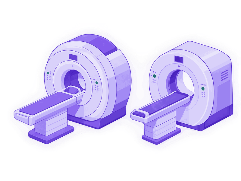
How an MRI differs from a CT
| MRI | CT | |
|---|---|---|
| What it shows | discs, nerves, spinal cord, ligaments, oedema | bone, fractures, tissue density |
| When it is ordered | pain in the leg or arm, numbness, weakness | injury, suspected fracture |
| Duration | 20–40 minutes | 5–10 minutes |
| Radiation | none | yes, X-rays |
| Limitations | pacemaker, metal in the body, claustrophobia | pregnancy |
In short: MRI shows the soft, CT shows the hard. A herniation, a compressed root, infection and a tumour are seen on an MRI. A fracture, bony overgrowth and the density of a vertebra, on a CT.
So for pain radiating into the leg you need an MRI. And after a fall from standing height in someone with osteoporosis, a CT.
Sometimes both are needed: when planning an operation for stenosis, for example, it matters to see both the soft tissue and the bony overgrowth.
Which part of the spine to scan
What to scan is decided by the doctor, not chosen by the patient: I will tell you at the visit or over the phone once you describe what hurts and how. Guessing is the second most common source of wasted money: people scan where it hurts most, when what should be scanned is where the nerve comes from.
Below, for reference, so the logic is clear. It is not a guide to prescribing for yourself, but an explanation of why leg pain means looking at the lower back.
- Pain and numbness in the leg - the lumbar spine.
- Pain and numbness in the arm and fingers - the cervical spine.
- Band-like pain around the ribs - the thoracic spine.
- Low back pain without anything shooting into the leg - lumbar, though in the first weeks a scan is often not needed at all.
- Numb little and ring fingers - the problem may not be in the spine at all but in the ulnar nerve. There a nerve conduction study is needed, not an MRI.
Scanning "the whole spine" is usually unnecessary: it takes longer and is no more accurate.
Do not decide on the part of the spine yourself. Describe what hurts and how when you book, and I will tell you which study of which part to have - or that it is not needed before the visit at all.
Are your scans out of date?
Two requirements that most people do not know about.
The machine must be 1.5 Tesla or stronger. Images from low-field scanners can be so uninformative that no decision about surgery can be made from them. The field strength is stated in the report.
No more than six months old. The picture changes: a herniation can shrink on its own or grow. A decision about surgery cannot be made from a two-year-old scan.
What to bring the scans on
A disc or a flash drive. At the consultation I load the study onto a computer and go through it myself, slice by slice; a report is no substitute for that. So bring the medium, not the paper - a disc or a flash drive with the study recorded on it. Nine times out of ten an imaging centre hands out a disc.
Take the report too, but it is secondary. It is a radiologist's opinion of what they saw. It is useful because it states the field strength and the date, but a decision about surgery is made from the images themselves.
What does not work. A phone photograph of a screen, a photo of printed film, a screenshot from a messenger. Nothing can be measured from those. Film itself is workable but worse than a disc: it cannot be scrolled through slice by slice.
If the disc is lost, go back to the centre where the study was done and ask them to burn another one. They keep the studies; it takes a couple of minutes.
A tip
If you are having an MRI for the first time, ask at once for the study on a disc or a flash drive, not just a printed report. It costs nothing and saves a second trip to the imaging centre.
What to do next
If you have the scans, book a visit. If you do not, call and I will tell you which study is needed and whether it is needed before the visit at all. Sometimes in the first weeks of pain no scan is required and it makes more sense to start treatment.